CMDCMiracle
심혈관 대사질환센터
Cardiovascular and Metabolic Disease Center
Mitochondrial Research Affinity Collaboration-Laboratories & Engineering
Facility
Equipment Protocol
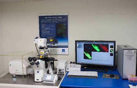
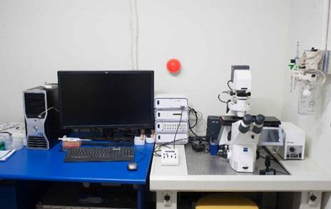
Imaging devices of the Biological Signal
- Study of the expression level of the protein
- Study of the position of the tissue or cell
- Physiological signal of cells: Measurement of calcium, sodium, and ROS
- Physiological signal of mitochondria: Membrane voltage, ROS, calcium signal detection
Our Research
- Changes in the calcium signal of Cardiomyocytes
- Changes in the calcium signal of Cancer cells
- Changes in the membrane voltage, calcium and ROS of Mitochondria
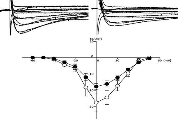
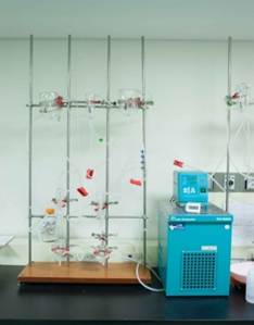
Patch Clamp
- Understanding of the function of ion channels
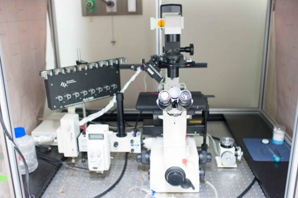
Our Research
- Changes in the Ion channel(Ca2+, K+, Na+ channel) of cardiomyocytes
- Ion movement in the cardiomyocyte
- Function and concentration of the ion
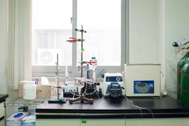
Organ Bath
- Evaluation of contraction / relaxation ability of the blood vessel
- Evaluation of contraction / relaxation ability of the blood vessels by drug
Our Research
- Evaluation of the contraction and relaxation ability in accordance with the anatomical position of the blood vessel
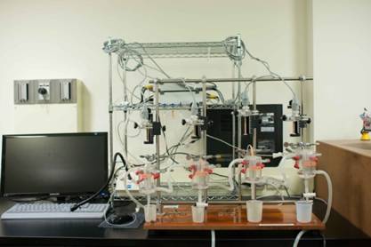
Langendorff System for Mouse and rat mode; constant volume
- Measurement of implemented pressure of the left ventricle of the heart
- Measurement of the pressure in the right ventricle of the heart
- Evaluation of left ventricular function of the heart
Our Research
- Evaluation of the Cardiac toxicity of Drug
- Evaluation of the cardioprotective effect about ischemia/reperfusion
- Identification of a signal transduction pathway associated with a contractile force of the heart
- Separation of cardiomyocyte
Echocardiography & Electrocardiogram (ECG)
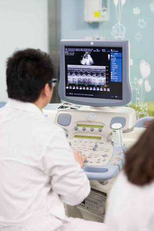
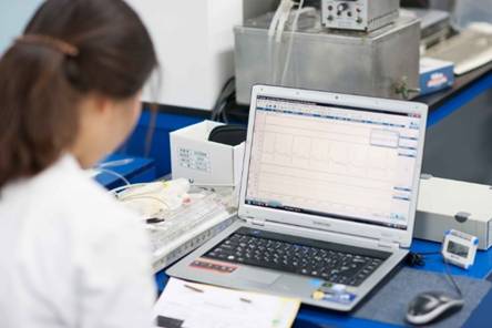
Our Research
- Construction of a therapeutic agent through the functional evaluation of the heart and vessels