To investigate tumors, pathologists currently rely on labor-intensive microscopic examination, using century-old cell-staining methods that can take days to complete and may give false readings. A lightning-fast laser technique has provided laboratory demonstrations of accurate, real-time, high-throughput identification of liver tumor cells at their earliest stages, and without invasive chemical reagents.
The technique generates a laser beam in single human cells pumped from a flask through tiny microchannels. The beam is altered by what it encounters. These changes, registered by an imaging spectrometer, instantly identify cancer-modified mitochondria in cells gone wrong. Mitochondria are known as the power pack of cells, energizing them like batteries do flashlight bulbs.
There are hundreds of mitochondria, sometimes thousands, in a cell. To see them in the old way requires a time-consuming process like fluorescent tagging or a chemical reagent.
To rapidly assess the health of a single mammalian cell, the key discovery was the elucidation of biophotonic differences in normal and cancer cells by using intracellular mitochondria as biomarkers for disease. This technique holds promise for detecting cancer at a very early stage and could nearly eliminate delays in diagnosis and treatment. It measures changes in the cell architecture, especially those arising from alterations in protein density, cytoskeleton shape, and distribution of mitochondria changes that occur when a cell becomes cancerous
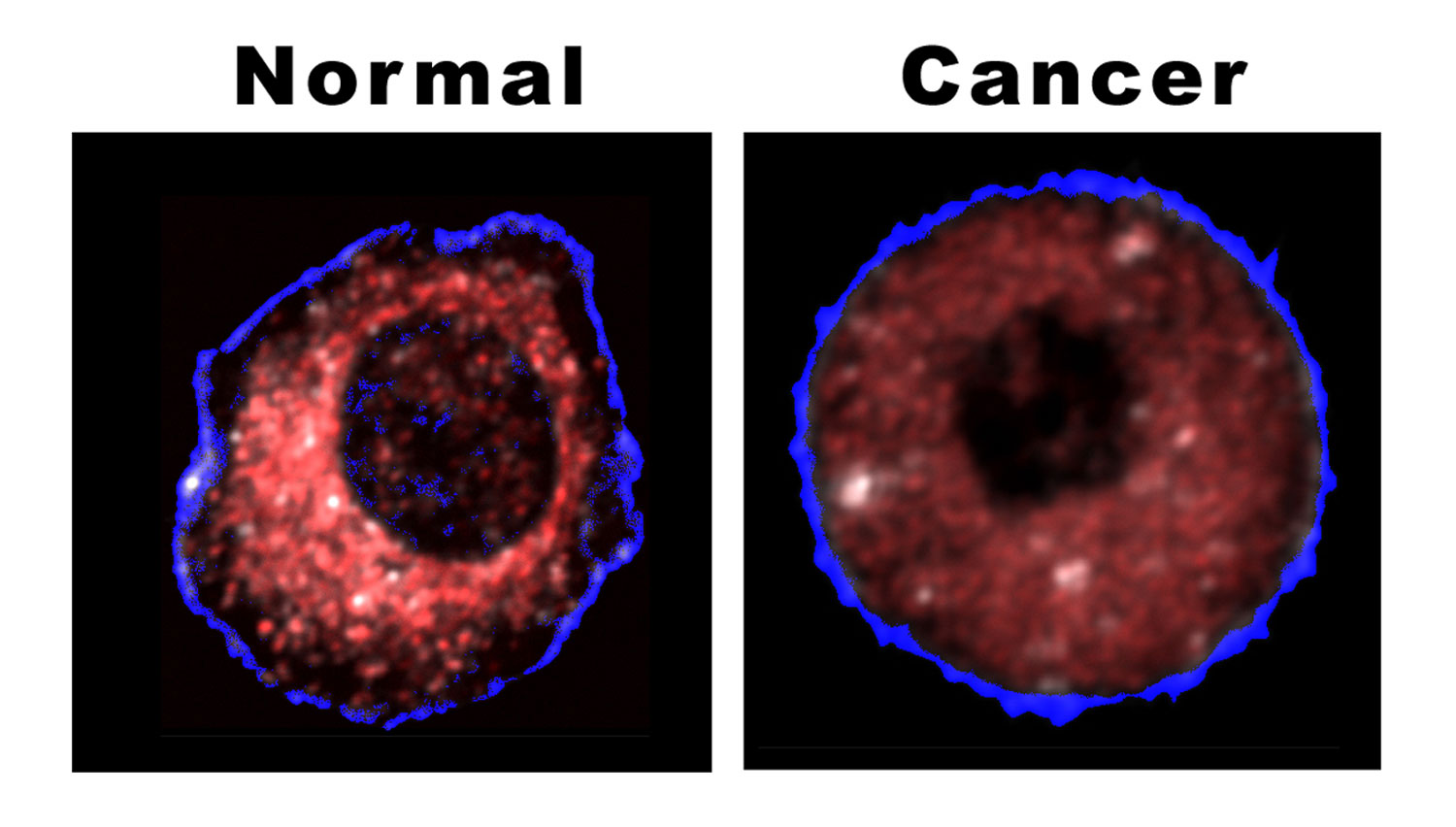
Image attached:
proof positive - The difference between a normal and cancerous liver cell is shown clearly by the location of mitochondria, as revealed by Sandia's biocavity laser. The healthy cell shows very few mitochondria near the outer cell wall; they cluster densely (red coloration) as they approach the cell's nucleous (depicted here as the black central hole). In the cancerous cell, the mitochondria are spread throughout the cell, do not cluster, and under the same lighting produce a more subdued effect.


